Introducción
The sacroiliac joint (SIJ) is a diarthrodial joint with innervation that has been the subject of debate in medical literature. Dr. Carlos Pozzoni et al. suggest that the sacroiliac joint is innervated by the posterior branches of the first two sacral nerves, as well as by the superior gluteal nerve, the sacral plexus, and the obturator nerve, which is a branch of the lumbar plexus.
One of the most common causes of low back pain is associated with sacroiliac joint pain, with a frequency of between 10 and 27 % (1). Sacroiliac pain is associated with risk factors such as gait disturbance, shortening of the lower limbs, obesity, pregnancy, spondyloarthropathies, and connective tissue diseases (2). Sacroiliac joint arthropathy can exist on its own, but it is commonly part of a complex pain syndrome such as disc degeneration, facet arthrosis, narrow canal, and post-laminectomy syndrome, etc. Spinal surgery, especially fixation in the lumbar region, is a predisposing factor for sacroiliac dysfunction syndrome (3).
Anatomy
Understanding anatomy is crucial for any interventionalist. As mentioned previously, there has been much debate about the innervation of the SIJ (sacroiliac joint). The anatomy is summarized in Figure 1. The nerves that innervate the posterior capsule result from a combination of the lateral branches of the primary sacral roots that run from S1 to S3 with a small contribution from S4, L4, and L5. (4)
Roberts et al. concluded the following in a cadaveric study: The posterior aspect of the SIJ was found to be innervated by a fine nerve plexus, the PSN (posterior sacral plexus or posterior sacral network), formed by the lateral branches of the posterior branches of L5-S4 (Fig. 1). The lateral branches of S1 and S2 contributed to the PSN in 100 % of the samples, S3 in 88 %, L5 in 8 %, and S4 in 4 %. The lateral branches and the PSN extended along a continuous plane at the level of the periosteum medially and on the posterior surface of the sacroiliac ligament laterally. Between the posterior sacral foramina and the lateral sacral crest, the lateral branches and the medial part of the PSN lie on the posterior aspect of the sacrum at the periosteal level and deep within the posterior short sacroiliac ligament. As the nerves of the PSN ran laterally from the lateral sacral crest, they lay down in the interosseous sacroiliac ligament and emitted branches that penetrated the ligament to innervate the SIJ. This lateral part of the PSN is lodged deep in the long posterior sacroiliac and sacrotuberous ligaments (4).
Consistent with previous studies, the current study found that the posterior aspect of the SIJ was innervated by the PSN, formed by the lateral branches of S1-S3 in most specimens, with S4 contributions being highly variable. In the current study, S4 innervated the SIJ in 1 specimen, whereas in the previous literature, in 2 of 7 studies, no contribution from S4 was found, and in the remaining studies, its contribution ranged from 13 % to 100 %. As in previous studies, in the current study, the contribution of the lateral branch of L5 was found to be inconsistent (2/7 studies and 8 % of samples, respectively). Although the number of lateral branches at each level varied from 1 to 3, they were found to emerge from the superolateral and inferolateral quadrants of the posterior surface of the sacrum, coinciding with the results of Yin et al., who reported that the branches emerged from the 2 to 6 o’clock position on the right or the 6 to 10 o’clock position on the left. One thing that remained constant was their location in the PSN just above the lateral sacral crest. This is important because denervation techniques should focus on the lateral sacral crest (4) (Figures 1 and 2).
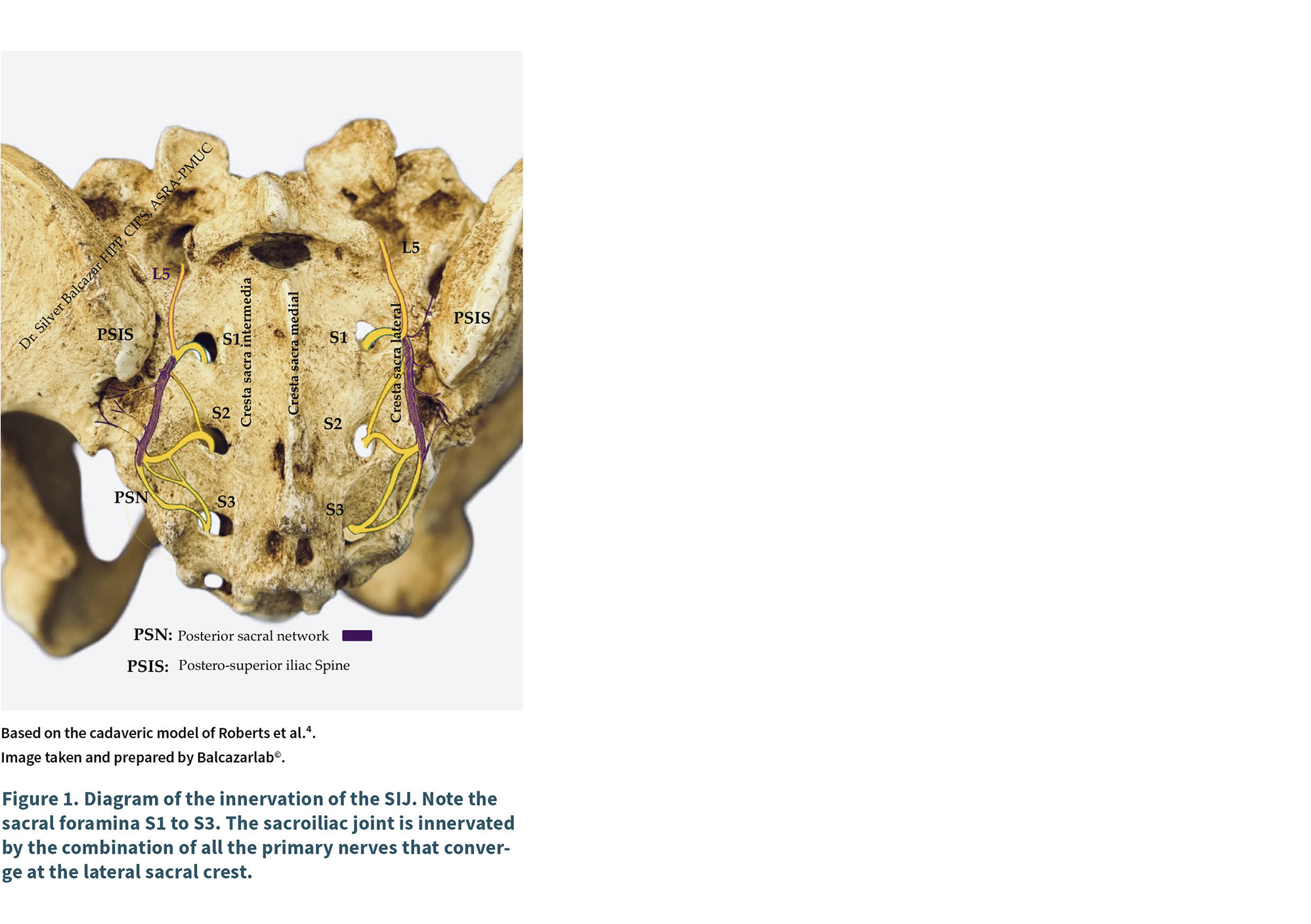
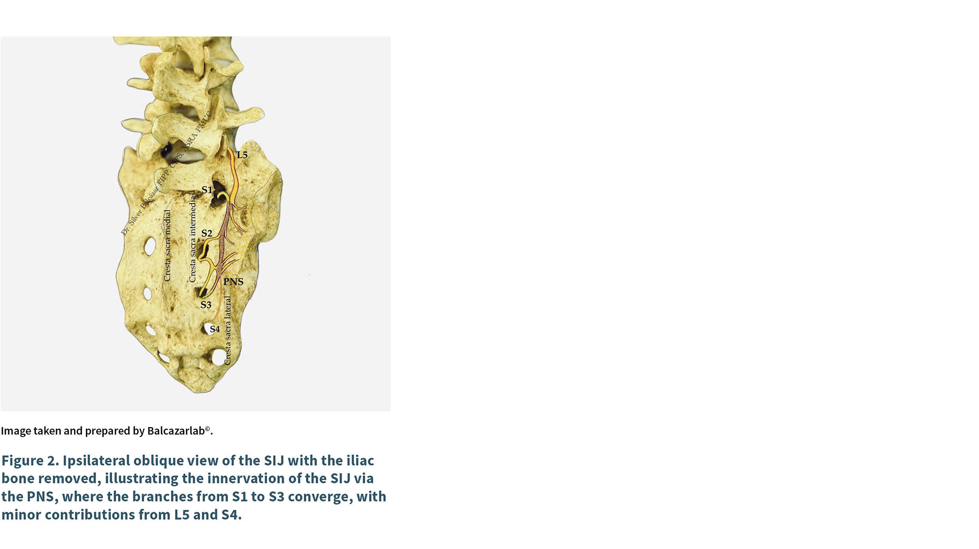
The lateral sacral crest is your friend, don’t forget to always keep it in mind
Traditionally, radiofrequency (RF) techniques have been used for SIJ denervation, including modalities such as conventional, bipolar, pulsed, and cooled RF. The results of ablation of the lateral sacral branches from S1 to S3 using bipolar radiofrequency have been evaluated, with significant pain improvement at 12 months. Wright also comments that to achieve complete denervation, the medial branch of L4 and L5 must be performed (5). It is important to note that until a few years ago, SIJ denervation was performed using fluoroscopy. However, anatomical studies by Roberts et al. have shown that ASI innervation is achieved through a plexus located on the lateral sacral crest, providing a starting point for identifying this structure, theoretically by ultrasound (4). Eldon et al. performed denervation of the sacroiliac joint using conventional radiofrequency with bipolar modality on the lateral sacral crest guided by ultrasound. In 31 patients studied, a significant reduction in pain was observed at 9 months. In this study, the ultrasound and fluoroscopy techniques were compared, with no significant differences found in either group at the 2-month follow-up. Preliminary results suggest that RF ablation of the SIJ can be performed under ultrasound guidance (6). Ablative treatments focus on conventional radiofrequency thermocoagulation, where the ablation temperature is above 45 °C, usually 80 °C, and are performed in a monopolar, bipolar, or tripolar manner, covering from S1 to S3 on the lateral sacral crest. RF ablation has been in use for decades and has proven to be effective in pain control. However, it destroys the nerve and surrounding tissue, with the possibility of neuroma formation. Furthermore, reinnervation occurs, but it is usually erratic and does not match the innervation pattern prior to destruction, thus reducing the likelihood of response with a second or third ablation. Cryoneuroablation has emerged as a promising alternative, with benefits such as a lower risk of neuromas because the effect is axonotmesis, preserving the axon and allowing nerve regeneration and more diffuse denervation by axonotmesis (7).
Cryoanalgesia using cryoneuroablation is a technique that dates back to 1961 when Cooper introduced a device that used liquid nitrogen. Current devices perform an axonotmesis-type lesion at a temperature between -20 and -100 °C. Current data show that it is effective in relieving pain for 6 to 12 months and also improves the functional capacity of patients with chronic non-cancer pain (8,9). Cryodenervation of the sacroiliac joint has been performed successfully, and its potential has been described since 2003 (7). Bellinger et al. (2015) published a series of cases evaluating the efficacy of cryoanalgesia in chronic pelvic pain, including patients with sacroiliac dysfunction, reporting a significant improvement in 70% of cases that lasted at least 9 months (10). Das in 2023 compared SIJ cryodenervation vs. the use of intra-articular corticosteroids, concluding that cryodenervation is superior to corticosteroids in pain relief (11).
Among the advantages of cryodenervation for the management of sacroiliac pain, we can mention: controlled nerve damage without permanent destruction of the endoneurium, lower risk of neuromas, less post-procedure pain, reproducible technique, and adaptable to image guidance (7).
SIJ cryoablation is feasible and safe, and ultrasound guidance is effective in this particular technique, allowing the combination of both to achieve the clinical result of pain reduction via ultrasound.
Case presentation
This is a case study of a 79-year-old female patient with no history of inflammatory arthritis who attended the clinic complaining of pain in the gluteal and sacral regions that had been present for six months, with a pain intensity of NRS 5-6/10, ODI (Oswestry Disability Index) of 38, Roland-Morris Disability Questionnaire (RMDQ) of 12 points, with intermittent and minimal improvement with NSAIDs. Pain worsened with prolonged standing and improved partially with rest. Minimal response to physical therapy.
Physical examination showed pain on palpation of the right sacroiliac joint and positive clinical tests for sacroiliac dysfunction (Patrick’s test, distraction, compression, and Gaeslen’s test). Imaging studies (X-ray and MRI) were performed, showing no signs of unilateral sacroiliac inflammation, but the X-ray showed signs consistent with SI joint sclerosis. Laboratory studies ruled out rheumatological pathologies. A diagnostic block was performed, which was positive, and the patient was admitted for ultrasound-guided sacroiliac joint cryodenervation.
Ultrasound-guided cryoablation sacroiliac denervation technique.
Step-by-step description
Under non-invasive monitoring, in the prone position, with local anesthesia and sterile technique, ultrasound-guided cryoneurolysis is performed using the following equipment: Metrum Cryoplex© cryosurgery generator. Cryoprobe 1.8 mm, 5 mm tip, gas used in this case: nitrous oxide, Sonosite PX ultrasound machine with 5 MHz convex transducer. EVA © ultrasound cover with sterile gel, 14 G introducer. Under sterile technique, ultrasound scanning is initiated with a 5 MHz convex transducer.
— Step 1: it is placed in a transverse position over the sacral horns to identify the sacral hiatus.
— Step 2: move toward the cephalic direction, in the same transverse position, until the sacral horns are lost and the medial sacral crest is identified as a hyperechoic image with acoustic shadowing, central relief, and two hyperechoic lines in each position.
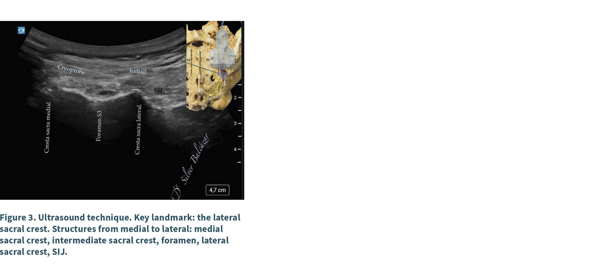
— Step 3: the transducer is then moved laterally to identify: the middle sacral crest, the intermediate sacral crest, the S3 foramen, and the lateral sacral crest (target area).
— Step 4: once the lateral sacral crest is identified at the S3 level, a 14 G introducer is inserted, in a medial to lateral plane (3 cm from the transducer to improve visualization), and the cryoprobe is accessed through the introducer until it contacts the lateral sacral crest.
— Step 5: sensory (50 Hz, maximum 1 volt) and motor (2 Hz, maximum 2 volts) checks are performed, and two cycles of lesions are performed at -88 degrees Celsius for 2 minutes with 30 seconds of defrost time between each lesion (Figure 3).
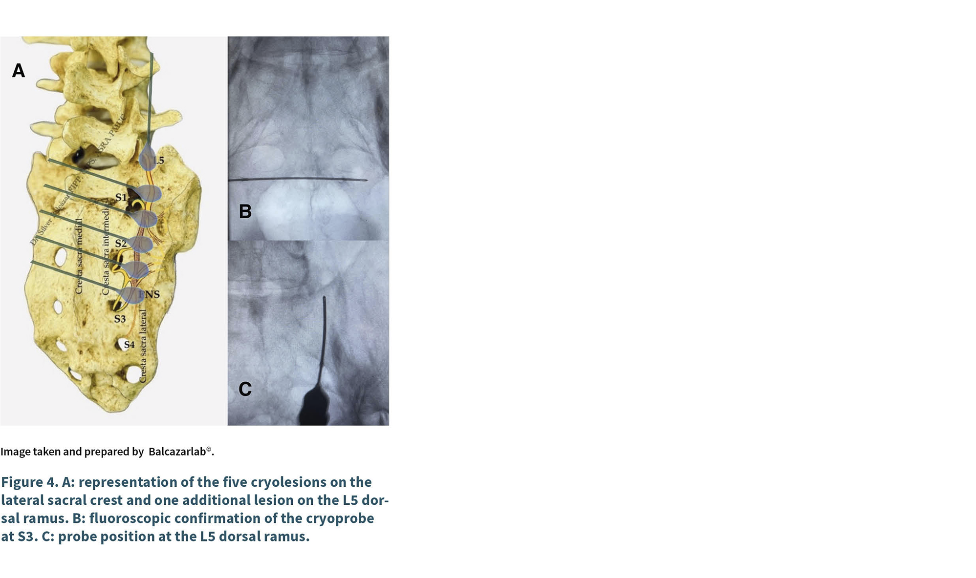
— Step 6: this process is repeated at a distance of 1 cm cephalad, just at the level of S3, S2, S1, and with an additional lesion between S2 and S1; all points are just above the lateral sacral crest, as shown in Figure 3.
A total of 5 ablation points and 2 cryoablation cycles are considered at each point with 30 seconds of defrosting between each lesion, all on the PNS on the lateral sacral crest, covering a length of at least 4 cm cephalocaudally on the lateral sacral crest. An additional lesion is made in the L5 dorsal branch, for a total of 6 cryoablation points. For illustrative purposes, each approach is checked with fluoroscopy to confirm that the ultrasound-guided placement is as accurate as the fluoroscopic approach (Figure 4).
The patient is discharged from the operating room to the post-surgical recovery room and, one hour later, is discharged home with follow-up by telephone 15 days after the procedure and in person 1 month and 3 months after the procedure. In the first 15 days, an NRS of 3/10 and an ODI of 16 are reported, which remain the same at 3 and 6 months. It is important to note that the difference in ODI (initial ODI - final ODI = 22 points), RMDQ 3 points at 6 months, with a reduction in pain medication and improvement in activities of daily living.
Discussion
Sacroiliac pain can be caused by: joint dysfunction, trauma, arthritis, or biomechanical alterations (3). The most common sites of pain are: the sacroiliac region (94 %), followed by the lower lumbar region (72 %), the lower extremities (50 %), the groin area (14 %), the upper lumbar region (6 %), and the abdomen (2 %). The innervation of the joint was explained in the introduction; therefore, the target nerves are the lateral branches of S1-S3 and the dorsal branch of L5, which is where denervation techniques are focused.
Sacroiliac denervation by ultrasound-guided cryoanalgesia is an emerging approach to the treatment of chronic sacroiliac joint pain, although specific evidence on this combined technique (cryoablation + ultrasound) is still limited. Das G 2023, (11) published a study comparing sacroiliac cryodenervation with the use of intra-articular corticosteroids, finding significant pain improvement in the cryoanalgesia group. This study performed denervation using ultrasound guidance, but only considered three targets (S1 to S3), using the lateral sacral crest approach. This is the only report on the use of this technique considering only the sacral roots S1 to S3, unlike our case report, which also considers the dorsal branch L5, which, as demonstrated in cadaver studies, contributes to innervation (4).
In this case, ultrasound-guided cryodenervation was performed on the dorsal branch of L5 and the lateral branches of S1-S3, taking into account five targets separated by 10 mm, with the aim of obtaining a larger lesion field (12 mm x 11 mm iceball) and making the blocking of pain transmission more efficient. Unlike radiofrequency thermoablation, cryodenervation does not generate heat, nor does it destroy the endoneurium or surrounding tissue. This translates into reduced postoperative pain, in addition to the maximum peak therapeutic effect observed at 6 days (4), without pain from neuritis and unlike RF, which usually takes up to 21 days for the clinical effect to appear, also considering that there is a risk of post-procedure pain. This makes cryoanalgesia a safe, effective, tolerable strategy with less pain and an early peak effect, in addition to generating much larger surface lesions compared to RF, which usually requires up to 6 or 7 cannulas for a lesion only 2 to 4 mm wide. Compared to cryoanalgesia, a lesion of more than 1 cm is achieved, which improves denervation compared to smaller lesions.
Ultrasound guidance is an alternative to fluoroscopy or tomography, offering real-time visualization without radiation and allowing the identification of nearby vascular/nerve structures. Always taking into account parameters such as the depth of the sacroiliac joint and bone interference that can hinder visualization, requiring operator experience, in addition to no radiation exposure, which means lower economic cost and greater feasibility. It is also accessible to specialties that do not have access to hemodynamics rooms.
Currently, studies are limited: there are more publications on radiofrequency (RF) or diagnostic blocks for sacroiliac pain. Cryoablation in this area is less well documented, although it has been explored in other joints such as the knee, shoulder, and ankle (12,13). The standard for interventional procedures on the spine is usually fluoroscopy. In the sacral spine, there are some advantages to performing procedures using ultrasound, for example, the structures are more superficial than in the lumbar region, and the neural and vascular structures are less exposed compared to the cervical spine. In addition, the bone offers an easily visualized target in the sacral approach, and it is usually feasible to identify the structures at this level. Previous studies (11) have successfully performed the ultrasound approach for ASI joint denervation with effective results. In addition, studies have been conducted on conventional radiofrequency ablation guided by ultrasound and compared with fluoroscopy, demonstrating not only that ultrasound is comparable and accurate, but also effective in the long term (6). Important limitations of this case report include the volume of patients, the fact that ultrasound often has limitations in overweight or obese patients, and the operator-dependent nature of ultrasound. Another important limitation is the long-term follow-up in this report.
Further studies are needed that not only compare the use of cryoanalgesia and radiofrequency, but also the use of ultrasound and fluoroscopy, as well as controlled clinical trials; however, this report is a good start to open up the field of research in this area.
Conclusion
Sacroiliac joint denervation by ultrasound-guided cryoanalgesia is technically feasible and promising, but its clinical adoption requires further study.
Conflict of interest
None.
Funding
None.
references
1. Pozzoni C, Sivanto MV, Rosenfeld N, Fandos L, Hlawaczeck C. Síndrome de disfunción sacroiliaca. Rev Arg Anest. 2001;59(2):124-8.
2. Sayed D, Deer TR, Tieppo Francio V, Lam CM, Sochacki K, Hussain N, et al. American Society of Pain and Neuroscience Best Practice (ASPN) Guideline for the Treatment of Sacroiliac Disorders. J Pain Res. 2024;17:1601-38.
3. Raj MA, Ampat G, Varacallo MA. Sacroiliac Joint Pain. En: StatPearls [Internet] [Internet]. StatPearls Publishing; 2023 [citado 20 de marzo de 2025]. Disponible en: https://www.ncbi.nlm.nih.gov/books/NBK470299/
4. Roberts SL, Burnham RS, Ravichandiran K, Agur AM, Loh EY. Cadaveric study of sacroiliac joint innervation: implications for diagnostic blocks and radiofrequency ablation. Reg Anesth Pain Med. 2014;39(6):456-64.
5. Wright RE, Block JE. Radiofrequency Neurotomy for Sacroiliac Joint Pain: Twelve Month Outcomes and Comparison Between Two Techniques. Med Res Arch. 2023;11(12). DOI: 10.18103/mra.v11i12.4978.
6. Loh E, Agur AM, Burnham RS. Ultrasound-guided radiofrequency Ablation for SI joint pain:An observational study. Interv Pain Med. 2022;1(3):100118.
7. Trescot AM. Cryoanalgesia in interventional pain management. Pain Physician. 2003;6(3):345-60.
8. Law L, Rayi A, Hendrix JM, Derian A. Cryoanalgesia. En: StatPearls [Internet]. Treasure Island (FL): StatPearls Publishing; 2025 Jan-. Disponible en: https://www.ncbi.nlm.nih.gov/books/NBK482123/
9. Goyal S, Kumar A, Sharma RS, Goyal D, Singh GK. Efficacy of cryoneurolysis in the management of chronic non-cancer pain: A systematic review and meta-analysis. Indian J Anaesth. 2022;66(7):485-97.
10. Bellini M, Barbieri M. Percutaneous cryoanalgesia in pain management: a case-series. Anaesthesiol Intensive Ther. 2015;47(4):333-5.
11. Das G, Das S, Sahoo R, Shreyas S, Kanthi B, Sharma VS. Efficacy of cryoneurolysis versus intra-articular steroid in sacroiliac joint pain: A retrospective, case-control study. Indian J Anaesth. 2023;67(11):1004-8.
12. Balcázar de León S, Carvajal Durán LI. Ultrasound-guided cryodenervation of the ankle joint in osteoarthritis. Case report and a novel percutaneous technique. MPJ. 2025;5:19-26. DOI: 10.20986/mpj.2025.1089/2025.
13. Balcazar de León S, Castro Guillén GN, Altamira García J, Vázquez Reyes JM. Criodenervación de complejo articular glenohumeral y acromioclavicular guiada por ultrasonido en hombro doloroso crónico. Análisis retrospectivo. MPJ. 2025;5:102-12. DOI: 10.20986/mpj.2025.1098/2025.